|
 |
Research Project P8:
Tracking Stem Cell Behavior in Microwells
Research Project Goal
The goal of this project is to use time-lapse microscopy to find, count and track stem cells over time. Individual cells are confined within an array of single microwells and over time the cell divides multiple times to yield a population of cells within each microwell. In this project, we want to use time-lapse microscopy images to determine the number of cells per well over time, record physical properties such as size, and also estimate the number of divisions.
Adult stem cells are the cells responsible for growth, repair and regeneration of tissues after birth and they are found in many different tissues, including brain, muscle, pancreas, skin, etc. These cells have great therapeutic potential because they can both self-renew, to make more stem cells, and differentiate, to form all the specialized cells of the tissue. One example of this therapeutic use is bone marrow transplants. Bone marrow contains hematopoetic stem cells, which can regenerate all the blood cell lineages. However, the use of stem cells for therapy is limited because there are very few stem cells in tissue. If the cells are plated in culture in hopes that they will divide and form more stem cells, the stem cells tend to lose their "stemmess," so they are no longer capable of regeneration.
In the Blau lab, we are working to create an environment, or artificial niche, that will enable the expansion of stem cells, such that when cells are plated in culture and divide, they will maintain their "stemmess." Towards this goal, we are studying single cells in microwells in order to understand division kinetics at a more fundamental level and testing the impact that different protein signaling has the kinetics of division. In previous work, we have manually counted the number of cells in thousands of different wells. This process is very time consuming! In addition these movies contain a HUGE amount of data and we are currently only utilizing a tiny portion of their potential. We are hoping to automate the cell counting process and proceed to more advanced analysis of this enormous volume of data.
Research Project Scope
Given time-lapse video of stem cells in microwells, find and count cells over time. Determine number of divisions and time at which division took place.
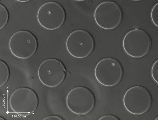 Day 1 (click on image to enlarge)
 Day 2 (click on image to enlarge)
 Day 3 (click on image to enlarge)
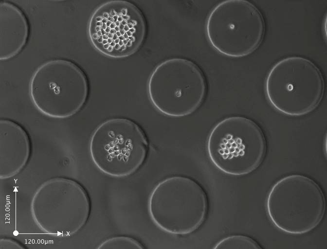 Day 4 (click on image to enlarge)
These are a few images from a time-lapse movie showing the cells dividing and increasing in number. The movie consists of images taken at regular intervals for 3-5 days.
Tasks
Find cells in microwells and count the total number of cells per well.
-
Record physical properties of cells, for example size, shape, mobility
- Estimate number of divisions per well
- Estimate time of divisions
- Plot data
Research Project Status
Ryan Matthew Smith
Stefan Mohler
Point of Contact
Karen Havenstrite: khaven@stanford.edu
Midterm Report
not yet submited
Final Report
not yet submitted
|
 |
|
|