Project Goal
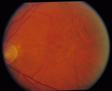
Retinal blood vessels are extremely important in many opthamalogical images. In some cases, they are of interest in and of themselves. In other cases, they simply obstruct the real visual target and must be ignored. In any case, accurate identification and segmentation of these structures is a very useful primitive.
Figure 1: Example retinal image.
Proper segmentation of blood vessels is also required for retinal image registration, a necessary first step for image-guided ocular surgery. The goal of this project is to come up with a fast and robust scheme to perform segmentation of blood vessels from retinal images.
Project Scope
This project involves interfacing with the Image Guidance Laboratories of Stanford's Department of Neurosurgery. The final product will be a blood vessel detector that should perform reasonably on even the most challenging retinal images. If there is time and interest, we could also try and make it work on mammography images (x-ray images of the breast), another, even more challenging, domain where blood vessel detection is important.
Tasks
The project will be accomplished through the following tasks.
- Task 1: Gain access to retinal image data from Stanford and from the Web.
- Task 2: Devise a fast and robust algorithm for the segmentation of blood vessels in retinal images.
- Task 3: Devise a way to validate your algorithm's results.
- (Extension): Test your algorithm on mammography images.
Project Status
Jennifer O'Meara, Teresa Miller and Paul EchevarriaPoint of Contact
Daniel RussakoffMidterm Report
submittedFinal Report
submitted


